Sunday, May 4, 2014

Bright or Dark? It’s your choice…

There’s a Malay saying, which stated that “ The young generation today is the leader in the future”.  It is wise to see this matter in larger context of the word leader. We should not narrow down the meaning of leader to only national or country leader. We are all leaders of the future. It’s how you plan and work for it. Do you want it bright or do you want it dark? It’s your own choice.

In the world of medical practice, who are the futures? Yes, you got the answer right it is the future doctors in the making now. Medical students? Yes it’s you, you and you. Wherever you are, whoever you are and whichever college you are in now, don’t forget you are the future of the world of medical practice and future of healthcare community. You are not there for no reason and for no expected outcomes. 

Plenty of question often comes to our mind as medical student as in : “ Am I competent enough when I graduated?”, “Is my medical school where I am studying now is in the same level of the other medical school?” , “There are lots of complained about the products of my medical school and what should I do now?” , “ DO I REALLY DESERVE TO BE A DOCTOR?”. These are some of the questions and dilemma in the hearts and mind of medical students. Gone are the days where you are in great joy to receive the offer letter from your medical school.  Where you are at least proud of yourself for a moment to be the crème de la crème to be accepted to medical school.

Special? Yes you are special to be in medical school but don’t forget the others in the other faculties deserve respects because they are special in their own way too as you are. Stepping your foot in the faculty doesn’t guarantee you anything without any effort done. People do fail exams, people do drop out from faculty and people do get extended for a year or more in the faculty. To be fair, in all faculties. The biggest mistake done is to consider yourself levels higher than the others to be in medical school.

Different from other medical school?  It is an undeniable fact that nowadays people tend to label according to medical school. Some are labeled as “now everybody can fly (pass)” , “useless graduate from ………..” ,  “ they let you pass the exam because you are the source of income of their school” and so on. It is heartbreaking to hear that from those who are from higher level in the practices. Don’t worry much, when there’s will , there’s always a ways out. If you think you are lacking behind in some part, make an effort to patch them out. Remember nothing is perfect in this world. There’s always pros and cons in each and everything. Some may look down on you because of your so called “incompetency” in clinical exposure. They may not realise your pre-clinical years were tough learning every basic sciences subject in and out like the post-graduates level.  Learning the tiniest muscle , vessels and nerves that may look unimportant. Still remember you asking yourself why should you learn some theoretical part which  seems have no role clinically? Sooner or later you will find out it will help you to understand things better and not to memorize  the clinical part. Some of us are really keen in knowing the clinical part but without knowing the reason of each and everything that happen. I still remember once my professor insisted me to present a case which I have just clerk without looking at what I have written down in the case note. Some medical school where people think that it is unsystematic just because they don’t really do piles of documentation. Too bad that’s a fact in some third world country. However in the other side , how many of us know the fact that due to lack of documentation, they, including the students were trained to know everything about the patients, not one but twenty or more by heart ? There is always a bright side of everything. It depends on how you are going to face it.

Your future is in your hand. It’s your choice to let it gloom or the other way round. One professor have told me before, treating a patient is not like answering a short question in an exam, you are treating the greatest creation of God. Now it’s up to you dear comrades, learning is not only in the classroom or during the obligatory ward rounds. Medical Science, which studies the greatest creation of God, is too broad and seems limitless. Get out of the box, gain more experiences even during medical school. It’s always never wrong to know more.  Bright or Dark? It’s your choice.

“Indeed, Allah will not change the condition of a people until they change what is in themselves”

Al-Quran 13:11


Monday, March 18, 2013

Pertolongan cemas (Bahagian 1)


Pertolongan cemas ialah bantuan segera atau rawatan kecemasan yang diberikan untuk orang yang tercedera sebelum mendapatkan bantuan atau perawat yang bertauliah. Ia biasanya terdiri dari satu siri teknik yang boleh dilakukan, walaupun tanpa latihan perubatan, oleh hampir semua orang dengan peralatan yang minimum. 


Objektif

  • Untuk menyelamatkan nyawa: Jika mangsa pengsan, pastikan laluan udara selesa dan pastikan mangsa bernafas. Sekiranya berlaku pendarahan, hentikan pendarahan secepat mungkin. Rawat kecederaan dengan betul dan kenali latar belakang mangsa (sejarah kesihatan seperti diabetes dan lain lagi) yang perlu perhatian istimewa.
  • Untuk mencegah luka menjadi lebih teruk: Alih mangsa dari tempat bahaya dan jangan buat mangsa banyak bergerak. Selesakan dan beri keyakinan pada mangsa serta beri perlindungan dari ketidak selesaan seperti kesejukan atau kepanasan atau keadaan yang basah.
  • Mendapat bantuan perubatan: Arah atau pinta rakan-rakan mengambil peti keselamatan berhampiran dan memanggil ambulans atau perawat bertauliah dengan sesegera mungkin. Pastikan menunggu bersama mangsa sehingga bantuan sampai.

Langkah Peringatan


Sebelum memberikan sebarang pertolongan cemas , sila pastikan keadaan sekeliling adalah selamat untuk anda. Hal ini bermaksud dalam proses memberi bantuan itu, keselamatan diri anda tidak terancam. Amalkan slogan "keselamatan diutamakan". Hal ini kerana kita tidak mahu menyebabkan diri anda tercedera dalam proses bantuan itu.


Contoh-contoh Keadaan serta Langkah-langkah Bantuan yang diberikan

Luka atau pendarahan

Luka yang kecil tidak memerlukan anda pergi ke hospital namun anda perlu mengelakkan luka tersebut daripada jangkitan kuman. Ikut langkah di bawah untuk memberikan bantuan kepada mangsa.

  1. Hentikan pendarahan : Luka kecil selalunya akan berhenti sendiri. Sekiranya pendarahan itu tidak berhenti,  berikan tekanan pada luka tersebut dengan kain atau pembalut yang bersih selama 20- 30 minit. Sekiranya boleh tinggikan aras pendarahan tersebut daripada aras jantung. Sekiranya pendarahan tidak berhenti lagi  hantar mangsa segera ke hospital.
  2. Bersihkan kawasan luka : Bersihkan kawasan luka dengan air yang bersih jangan gunakan sabun kerana sabun boleh menambah kesakitan pada kawasan luka. Bersihkan kawasan luka itu sehingga tiada kesan kotoran serta bahan asing. Sekiranya terdapat bahan asing seperti kaca jangan dibuang kaca itu sendiri, dapatkan bantuan pakar.
  3. Sapukan antiseptic : Sapukan cecair antiseptic di kawasan luka sekiranya ada. Sebaiknya gunakan bahan yang tidak memedihkan seperti iodine, elakkan menggunakan antiseptik seperti alkohol dan dettol.
  4. Balut luka : Balut luka tersebut dengan pembalut yang bersih, jangan terlalu ketat dan terlalu longgar
Pendarahan Hidung 

Pendarahan hidung merupakan keadaan yang biasa. Selalunya tidak merbahaya walaubagaimanapun ada beberapa kes pendarahan hidung disebabkan oleh kes yang perlu dirawat.

Langkah-langkah bantuan :

  1. Duduk tegak dan tundukkan kepala kehadapan. Jangan angkat kepala kerana perbuatan ini boleh menyebabkan darah itu masuk ke dalam mulut dan menyebabkan anda tertelan darah tersebut. Seterusnya boleh menganggu perut anda.
  2. Picit hidung anda untuk memberhentikan pendarahan untuk selama 5-10 minit. Bernafas melalui mulut.
  3. Elakkan pendarahan berulang selepas pendarahan berhenti dengan mengelakkan daripada memasukkan jari kedalam rongga hidung.
Dapatkan bantuan daripada pakar sekiranya :

  1. Pendarahan tidak berhenti selepas 20 minit
  2. Pendarahan disebabkan kecederaan seperti jatuh, kemalangan mahupun terkena tumbukan.
Melecur/Terbakar

Untuk memberi bantuan kepada mangsa yang mempunyai kesan melecur akibat terbakar di badan. Anda perlu mengetahui tahap Melecur mangsa tersebut.

Tahap yang Ringan 

Tahap pertama 

Biasanya tahap pertama hanya pada bahagian luar kulit.

  1. Kulitnya berwarna kemerah-merahan
  2. Berlakunya pembengkakan
  3. Selalunya mangsa merasa kesakitan di kawasan itu.
Tahap kedua

Tahap kedua kulit terbakar sehingga kawasan  yang dipanggil dermis.

  1. Wujudnya kesan lepuh (blister)
  2. Kulit nampak sangat merah
  3. Kesakitan yang teramat serta pembengkakan
Tahap yang Serius

Tahap ketiga

Kawasan yang terbakar teruk sehingga ke kawasan otot dan tulang. Berwarna hitam arang (hangus)


Langkah-langkah bantuan

Tahap ringan

  1. Sejukkan kawasan yang melecur atau terbakar dengan air. Alirkan air yang bersih berterusan di kawasan yang terbakar. Jangan gunakan air batu atau cecair lain seperti ubat gigi, mentega dan lain-lain
  2. Balut kawasan tersebut dengan pembalut yang bersih
  3. Dapatkan bantuan daripada pakar.
Tahap serius

  1. Dapatkan bantuan kecemasan melalui talian kecemasan
  2. Jangan tanggalkan pakaian mangsa di kawasan yang terbakar
  3. Jangan alirkan air
  4. Periksa nadi dan pernafasan. Sekiranya tiada berikan bantuan CPR (akan diterangkan di bahagian seterusnya kerana CPR memerlukan latihan dan penerangan yang lanjut)
  5. Tutup kawasan terbakar dengan  kain yang bersih.

Sekian sahaja untuk kali ini, dengan izin Allah, saya akan mengulas lebih lanjut mengenai pertolongan cemas seperti topik CPR, tulang patah dan lain-lain lagi dengan lebih mendalam. Hal ini kerana topik-topik sedemikian memerlukan penerangan yang teliti. Mohon maaf sekiranya ada kesilapan. Sekiranya ada sebarang kesalahan  atau pertanyaan diharap dapat terus hubungi kami.

Sekian Terima Kasih.

We Learn, We Share, We Care


Wednesday, January 16, 2013

Leptospirosis (Kencing Tikus)

Assalamualaikum.

Saban hari topik kencing tikus ataupun nama saintifiknya leptospirosis ini sering kedengaran terutamanya di tempat penulis menuntut. Ada sahabat kami telah pulang ke rahmatullah kerana penyakit ini.
Al-Fatihah.

Ramai yang bertanya apa sebenarnya penyakit ini dan mengapa boleh menyebabkan kematian. Oleh itu izinkan saya untuk mengupas secara ringkas mengenai penyakit ini.

Leptospirosis disebabkan oleh bakteria Leptospira spp. Bakteria ini menjangkiti manusia melalui air kencing haiwan yang mengandungi bakteria ini selagi mana air kencing itu masih dalan keadaan lembap. Walaupun tikus merupakan pembawa utama , haiwan lain seperti anjing, kambing dan lembu juga boleh menjadi pembawa sekiranya haiwan itu terjangkit dengan air kecing tikus yg bertidak sebagai pembawa utama.

Manusia boleh dijangkiti sekiranya termakan makanan yang dicemari oleh bakteria ini. Selain itu jangkitan juga boleh berlaku dengan sentuhan pada kulit.

Mereka yang selalu bergelumang dengan air tercemar merupakan golongan yang berisiko tinggi.

Tanda- tanda leptospirosis:

Demam
Sakit kepala
Kesejukan
Sakit otot
Muntah
Sakit bahagian abdomen
Cirit-birit
Kekuningan
Jangkitan selaput otak
Sentiasa penat
Kesukaran bernafas
Kehilangan pendengaran

Sekiranya anda berada dalam risiko tinggi serta mempunyai tanda-tanda bolehlah pergi ke pusat kesihatan yang terdekat.

Kita sebagai manusia hanya boleh merancang dan berusaha mengelakkan risiko namun ajal maut dan sakit itu semuanya ketentuan Allah SWT.

Sampai sini sahaja untuk kali ini. Andai ada persoalan boleh terus hubungi kami .
Maaf andai ada apa-apa kesilapan.


Monday, November 12, 2012

Appendicitis


Assalamualaikum WBT.

Hi everyone! Have you ever heard about APPENDICITIS?
I'm sure it is quite common to hear worldwide as it is the most common acute abdominal condition the surgeon is called on to treat. But, did we really know what is it all about?


Appendicitis & its pathophysiology.

  • Appendicitis is the inflammation of the appendix, which is either acute (most common) or chronic. It is associated with obstruction of the appendix, may be in the form of stool, foreign objects, tumor, or gallstone from the caecum, which enters the appendix and causing blockage
  • This stool will hardens, become a rock-like mass. When the blockage occurs, the bacteria will invade the wall of the appendix and causing inflammation.
  • Perforation or rupture of the appendix may occur if there is no treatment. This may lead to peritonitis, sepsis, and death. 
  • In Neuroimmune appendicitis:  Pain without acute inflammation, increase substance P / VIP, and non-inflammatory. 


How to diagnose?

      1. Laboratory:  - Leukocytosis with Lt. shift
                             - Total WBC count: < 10K/uL
                             - Absolute neutrophil count: < 6750/mL
                             - Hyponatremia
                             - Acidosis

      2. Radiography:  - X-rays (not specific, may shows air fluid)
                               - Calcified stone in appendiceal area
                               - CT scan and ultrasound (more accurate)

      3. Differential diagnosis:
              -Crohn's disease, Psoas abscess, Pyelonephritis, Pelvic abscess,
               ovarian/fallopian diseases, Cholecystitis, Intestinal perforation
               due to obstruction, or in male: Scrotum abscess, Hernia.


How to treat?

* Surgery : by removal of appendix by surgery OR
                 laparoscopic surgery ( less wound and risk, rapid healing)

* Fluid management is critical.


Symptoms & Scoring.

Apparently, all these things were quite complicated for non-medical practitioners to understand, so here it is. Alvorado scoring! It is usually used by the physician on the symptoms and scoring for the possibilities of appendicitis. But now you could self-diagnose yourself at home! (excluding the lab tests)  :)


You may also have


  • Dull pain near the navel or the upper abdomen that becomes sharp as it moves to the lower right abdomen. Or anywhere in the upper & lower abdomen, back, and rectum.
  • Loss of appetite
  • Abdominal swelling
  • Inability to pass gas
  • Constipation & diarrhea with gas
  • Painful urination
  • Severe cramps


So if you have most of the symptoms, quickly go get yourself checked!


We Learn, We Share, We Care

** Sorry for any inconvenience since this is my first post. :)


References: 
Robbins Basic Pathology (8th Edition)


Analysis of Symptoms (Endocrinology)

﷽

Analysis of symptoms in Endocrinology
Referring to how you analysis a symptom in the last post here are the common clinical features in endocrine disease.

Ask for:
  1. Weight Gain - Hypothyroidism, PCOS, Cushing Syndrome
  2. Weight Loss - Hyperthyroidism, DM, Adrenal Insufficiency
  3. Short stature - Constitutional, non-endoncrine systemic disease e.g. coeliac disease , GH deficiency
  4. Delayed Puberty - Constitutional, non-endocrine systemic disease e.g. hypothyroidism, hypopituitarism, primary gonadal failure
  5. Menstrual disturbance - PCOS, hyperprolactinaemia, thyroid  disfunction
  6. Diffuse neck swelling - Simple goitre, Graves' disease, Hashimoto's thyroiditis
  7. Excessive thirst - DM or Insipidus, hyperparathyroidism, Conn's syndrome
  8. Hirsutism - Idiopathic, PCOS, Cushing's Syndrome , congenital adrenal hyperplasia
  9. 'Funny Turns' -  Hypoglycaemia, phaeochromocytoma, neuroendocrine tumour
  10. Sweating -  Hyperthyroidism, hypogonadism, acromegaly , phaeochrmocytoma
  11. Resistant Hypertension - Conn's syndrome, Cushing's , phaeochromocytoma ,acromegaly , renal artery stenosis
  12. Erectile Dysfunction -  Primary/2ndry hypogonadism, DM, Non-endocrine systemic disease
  13. Muscle Weakness - Cushing's syndrome, hyperthyroidism, hyperparathyroidism, osteomalacia
  14. Bone fragility and fractures - Cushing's syndrome , hypogonadism, hyperthyroidism
  15. Altered facial appearance - Hypothyroidism, Cushing's Syndrome, acromegaly, PCOS

These are few common symptoms to be analysis in an endocrine case :)


References : Macleod's Clinical Examination 12th Edition

History Taking

Assalamualaikum WBT.

Hi guys long time since the last update from TAPMED. Today I would like to talk about history taking. History taking from patients is important to reach a good diagnosis. We can't deny that nowadays there are lots of high-tech work up or investigation that can bring you to a diagnosis. However looking back to years back where there are no such investigation, history taking plays a big role in reaching a diagnosis. So waste no time and lets begin to talk about history taking from patients.


First of all when you are with a patient, make him comfortable and introduce yourself. Tell him/her why you are here. Shake hands if possible try to make the patient know that you are eager to help him/her.

Then start with:

Personal History

Name : Get their full name , if in Egypt get three names like Ahmad Mohammed Ibrahim
Age : Some diseases are age related
Sex :  Some diseases are sex related
Occupation : To look for occupational disease
Residence : Some disease are endemic in specific places
Habit : Special habit of medical importance such as smoking, alcohol intake.
Menstrual History for women : Regularity, Days of Cycle , Amount of bleeding

Complaint (c/o)

Ask the patient what brings him to the hospital.
Make sure that you are writing it down in patients own word. Exclude medical terms
As short as possible (one complaint is enough)

e.g. don't write epixstasis , but write "complaining of nose bleed"

don't forget to write the duration. e.g. complaining of chest pain for 2 days

History of Present Illness

Do it in chronological order. Ask when he was last healthy (symptomless). Write in medical terms.
Then analysis the complaint.
Then ask for symptoms of the related systems then proceed for other systems.

For every symptoms analysis for

Onset/Course/Duration
Association
What will increase or decrease the symptom
Effect of TTT if there is any
Date of last attack

For pain ask for site, radiation and character of the pain.

This is the part where you are playing a big role. Don't let the patient lead you, but you should lead the conversation for this part.

Past Medical History

Ask for any disease such as DM, HPT, TB, I.H.D
Ask for it's duration, manifestation, investigation, ttt and complication
Ask for any surgical history when ,where(site) and out come. Don't forget to ask if any blood transfusion.
Ask also if patients is taking any drugs for treatment.

Family History
Ask for consanguinity, Similar conditions or any related chronic disease like DM/HPT/TB/I.H.D.


Wednesday, October 17, 2012

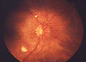
Diabetic Retinopathy (Short Notes)


Diabetic retinopathy,is retinopathy (damage to the retina) caused by complications of diabetes, which can eventually lead to blindness.

Classification

Non-proliferative :  Retinopathy, Maculopathy
Proliferative Retinopathy

Non-Proliferative Retinopathy


Manifestations:

Mild : At least one microaneurysm

Moderate : 

  • Extensive microaneurysm 
  • Intraretinal haemorrhages
  • Venous beading
  • Cotton wool spots
Severe :

  • Cotton wool spots
  • Venous beading
  • Intraretinal Microavascular Abnormalities (IRMA)


Nonproliferative Maculopathy


Manifested by focal or diffuse retinal thickening or edema due to breakdown of the inner blood retinal capillary endothelium causes leakage of fluid and plasma into surrounding of retina.

More common in type II diabetes which requires treatment once significant which is confirmed by any retinal thickening within 500 microns of the fovea, hard exudates withing 500 micron of the fovea with retinal thickening. Also retinal thickening greater than one disc diameter in size

It may due to schema which is characterized by macular edema, deep haemorrhages and little exudation.

Fluorescein angiography shows loss of retinal capillaries with enlargement of the foveal avascular zone.


Proliferative Retinopathy


It is the most severe complications of Diabetes 

It is characterised by NVD & NVE

Diagnosis


Diabetic retinopathy is detected during an eye examination that includes:
  • Visual acuity test: This test uses an eye chart to measure how well a person sees at various distances (i.e., visual acuity).

  • Pupil dilation: The eye care professional places drops into the eye to widen the pupil. This allows him or her to see more of the retina and look for signs of diabetic retinopathy. After the examination, close-up vision may remain blurred for several hours.

  • Ophthalmoscopy or fundus photography: Ophthalmoscopy is an examination of the retina in which the eye care professional: 

  1. looks through a slit lamp biomicroscope with a special magnifying lens that provides a narrow view of the retina.
  2. wearing a headset (indirect ophthalmoscope) with a bright light, looks through a special magnifying glass and gains a wide view of the retina. Hand-held ophthalmoscopy is insufficient to rule out significant and treatable diabetic retinopathy. Fundus photography generally recreate considerably larger areas of the fundus, and has the advantage of photo documentation for future reference, as well as availing the image to be examined by a specialist at another location and/or time.

  • Fundus Fluorescein angiography (FFA): This is an imaging technique which relies on the circulation of Fluorescein dye in the eye vasculature.

  • Optical coherence tomography (OCT): This is an optical imaging modality based upon interference, and analogous to ultrasound. It produces cross-sectional images of the retina (B-scans) which can be used to measure the thickness of the retina and to resolve its major layers, allowing the observation of swelling and or leakage.

  • Digital Retinal Screening Programs: Systematic programs for the early detection of eye disease including diabetic retinopathy are becoming more common, such as in the UK, where all people with diabetes mellitus are offered retinal screening at least annually. This involves digital image capture and transmission of the images to a digital reading center for evaluation and treatment referral. 

  • Slit Lamp Biomicroscopy Retinal Screening Programs: Systematic programs for the early detection of diabetic retinopathy using slit-lamp biomicroscopy. These exist either as a standalone scheme or as part of the Digital program where the digital photograph was considered to lack enough clarity for detection and/or diagnosis of any retinal abnormality.
The eye care professional will look at the retina for early signs of the disease, such as: 

  1. leaking blood vessels
  2. retinal swelling, such as macular edema
  3. pale, fatty deposits on the retina (exudates) – signs of leaking blood vessels
  4. damaged nerve tissue (neuropathy) 
  5. any changes in the blood vessels.

Should the doctor suspect macular edema, he or she may perform fluorescein angiography and sometimes OCT.

Treatment and Management

  1. Laser photocoagulation
  2. Modified Grid Laser photocoagulation
  3. Panretinal photocoagulation
  4. Intravitreal triamcinolone acetonide
  5. Intravitreal Anti-VEGF
  6. Vitrectomy


References:


  1. Vaughan & Asbury's General Opthalmolgy 17th Edition
  2. http://en.wikipedia.org/wiki/Diabetic_retinopathy


Diabetes Mellitus (Kencing Manis)


Artikel ringkas pertama yang bakal saya kongsikan adalah berkenaan Diabetes Mellitus ataupun lebih dikenali dalam kalangan masyarakat sebagai Kencing Manis. Jelas dan nyata tidak dapat dinafikan lagi bahawa penyakit ini merupakan salah satu daripada penyakit yang menjadi ancaman kepada masyarakat atas pelbagai sebab seperti  gaya hidup dan makanan.

Apa itu Diabetes Mellitus?

Diabetes Mellitus merupakan satu sindrom di mana kandungan gula dalam badan lebih tinggi dari paras normal  dan disebabkan oleh tiada insulin dihasilkan atau kurang insulin atau insulin tidak berkesan untuk mengawal paras kandungan gula dalam badan.

Paras Normal kandungan gula

Dalam keadaan berpuasa : 

Kurang daripada 110 mg/dL atau 6.1 mmol/dL

Dalam keadaan biasa :

Kurang daripada 140 mg/dL atau 7.8 mmol/dL

Paras disahkan Diabetes

Dalam keadaan berpuasa :

Melebihi  126 mg/dL atau 7.0 mmol/dL

Dalam keadaan biasa :

Melebihi 200 mg/dL atau 11.1 mmol/dL

*Paras di antara normal dan disahkan menunjukkan anda berisiko tinggi untuk menghidapi penyakit tersebut.

Adakah anda...........?

  1. Cepat merasa penat
  2. Selalu merasa dahaga
  3. Kerap ingin membuang air kecil
  4. Turun berat badan tanpa sebab
  5. Luka lambat sembuh
  6. Daya penglihatan semakin kurang
Andai anda mempunyai tanda-tanda seperti di atas, anda berisiko menghadapi penyakit kencing manis. Anda dinasihatkan untuk berjumpa dengan doktor untuk pemeriksaan lebih lanjut dan rawatan secepat mungkin andai anda disahkan menghadapi penyakit ini.

Kenapa diabetes perlu dikawal?

Diabetes perlu dikawal untuk mengelakkan komplikasi-komplikasi yang tidak diinginkan seperti :

  1. Kerosakan Mata (Penglihatan)
  2. Kerosakan buang pinggang
  3. Kerosakan urat saraf yang boleh membawa kepada ulser pada luka di kaki yang seterusnya menyebabkan kaki perlu dibedah ataupun dalam bahasa masyarakat "dibuang/dipotong"
  4. Atherosclerosis iaitu salur darah menjadi tidak kenyal boleh membawa kepada masalah salur darah tersumbat, masalah jantung , dan juga menghalang sumber darah ke kawasan tangan dan kaki.
  5. Menyebabkan risiko kepada penyakit tekanan darah tinggi.
  6. Menyebabkan risiko kepada peningkatan kandungan kolestrol dalam darah.
Elakkan daripada Diabetes!

Sesal dahulu pendapatan, sesal kemudian tiada gunanya.

  1. Kurangkan pengambilan gula dalam makanan dan minuman
  2. Bersenam
  3. Berhenti Merokok
  4. Jauhi arak 
  5. Pastikan berat badan dalam keadaan yang optimum


Setakat ini perkongsian pada kali ini, semoga perkongsian ini membawa manfaat kepada semua. InsyaAllah pihak TAPMED akan mengupas lebih lanjut dalam isu ini. Saya bagi pihak TAPMED ingin memohon maaf seandainya ada kesalahan dalam fakta , bahasa atau apa pun kekurangan dalam artikel ringkas ini.

Buah cempedak di luar pagar,
Ambil galah tolong jolokkan;
Saya budak baru belajar,
Kalau salah tolong tunjukkan.

Sekian sehingga berjumpa lagi.


We Learn, We Share , We Care